What is a Fetal Scan?
A fetal anomaly scan, also known as ultrasound or targeted ultrasound, is a specialised imaging procedure that provides a detailed examination of a developing fetus inside the womb.
Is a Fetal Anomaly Scan Compulsory During Pregnancy?
A fetal anomaly scan is performed between the 18th and 22nd week of pregnancy. Its purpose is to detect any structural abnormalities in the developing fetus. This non-invasive procedure uses sound waves to create detailed images of the body, allowing healthcare professionals to assess the baby’s growth and development.
Though the decision to go for this scan is personal, healthcare professionals strongly recommend this scan. Fetal anomaly scans detect abnormalities early, helping you make informed choices and take prompt medical attention when required.
When Do You Need It?
Prenatal care is the most essential aspect of pregnancy. This scan is performed around 18-22 weeks of pregnancy. This timing offers an accurate assessment to ensure the baby’s proper development inside the womb. You’ll also have ample time to make smart decisions when anomalies are detected.
Are Fetal Scans Safe?
Fetal scans are safe during pregnancy because ultrasound scans don’t involve radiation. Generally, low-risk pregnancies only require one to two ultrasounds during pregnancy. So, go ahead and see and feel your baby growing at each stage of the journey without holding back in fear.
There are 4 essential scans that are performed during each phase of pregnancy.
Types of Fetal Scans
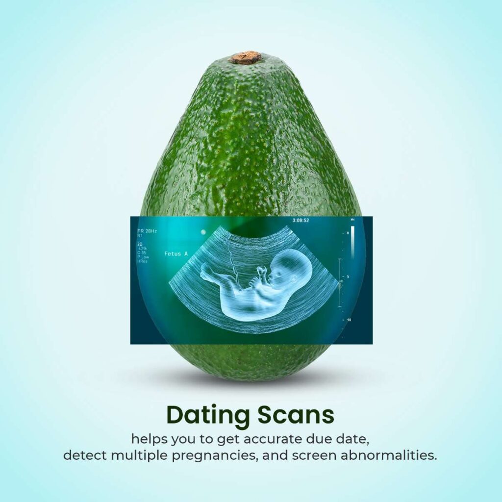
- Dating or early pregnancy scan
This is done before 12 weeks. This is the initial scan that confirms your pregnancy and due date.
- NT scan
Performed between the 12 and 14th week, an NT scan can detect any potential abnormalities that might occur in the future and also assess the structure of the fetus.
- Anomaly scan
An anomaly scan, taken around the 20th week, will allow you to examine the growth of the baby and its organs while also detecting any possibilities for chromosomal abnormalities that might cause conditions like Down’s syndrome.
- Growth scan
A growth or well-being scan is taken around 28 to 30 weeks and determines the baby’s full growth, fluid levels, blood vessels, blood flow, etc.
How is a Fetal Scan Done?
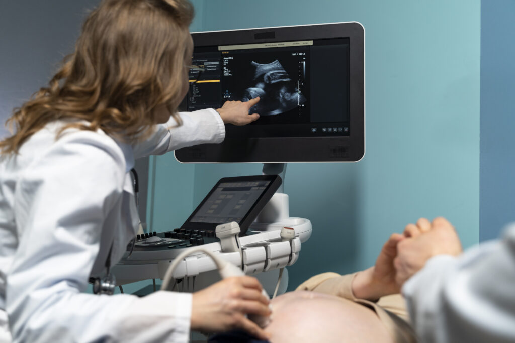
Having a full bladder during fetal scans is always advisable for better visualisation during your ultrasound. As you lie back on an examination table, a water-based gel will be applied to your abdomen, which helps in transmitting the sound waves through the ultrasound. A transducer is then moved across your belly, emitting and receiving sound waves to create images on the screen.
Amniocentesis
A thin needle is inserted into the amniotic sac through the abdomen to collect a small amount of amniotic samples for testing. This is performed on women with high-risk pregnancy chances with a birth defect.
Bottomline
Performing fetal scans allows you to enjoy risk-free and happy pregnancy moments, knowing the baby’s condition is safe inside the womb. Go ahead and take one step closer to safe pregnancy wellness